![HS-2700V [NEW] (from March 2020)](https://en.honda-el.co.jp/hubfs/images/product/ufile/thumbnail/medical/img_thum_medical_hs-2700v.png)
-
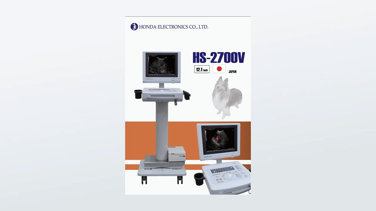
12. 1 inch high-resolution LCD.
-
Simple and easy-to-use keyboard design.
-
Sharp and smooth image display by H-res.
-
Large casters allow even women to move it easily.

Excellent ultrasound images
Our original image enhancement technology
H-res (Our Resolution Technology)
The development of ultrasound technology over the years, crystallized into the image enhancing technology as "H-res".
Optimum image can be achieved by adjusting the "H-res" parameter for each application and probe.
*Resolution visualizes delicate tissue structure in shallower region.
*Penetration visualizes good resolution in deeper organs.
*Boundary visualizes bones for observation.
*Clarity reduces the noise in blood vessel.
*Mild reduces the image enhancement effect.
*OFF has direct ultrasound image.
Kidney (cat)
Kidney (cat)
Gall bladder (dog)
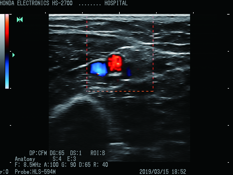
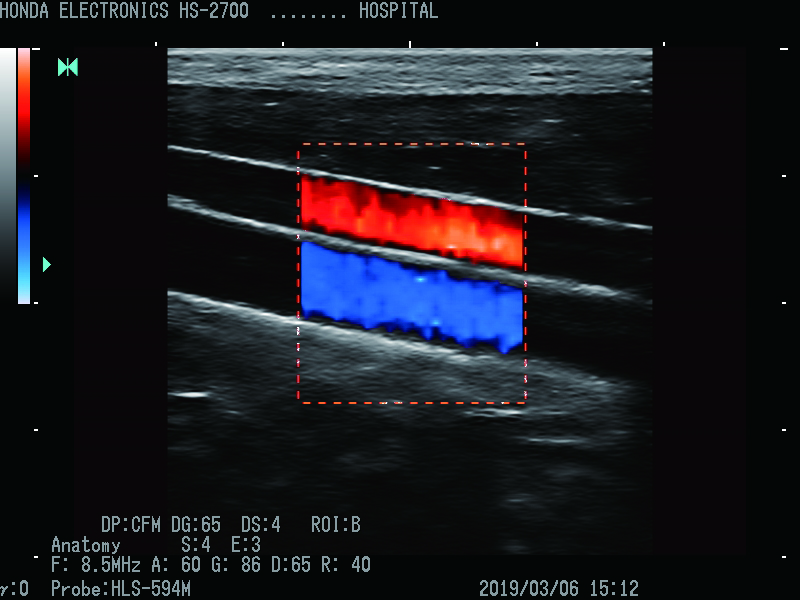
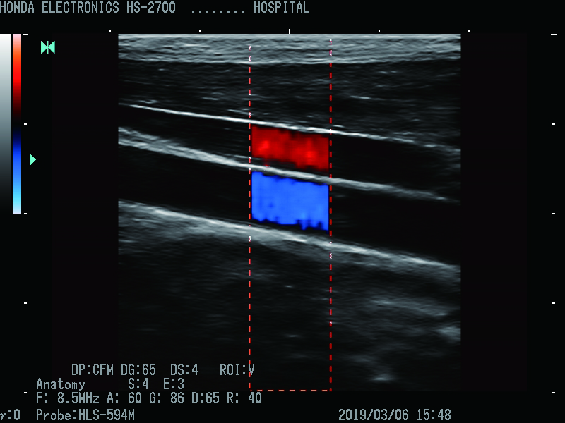
CFM(Color Flow Mapping)
Probe:HLS-594M
H-res:Anatomy
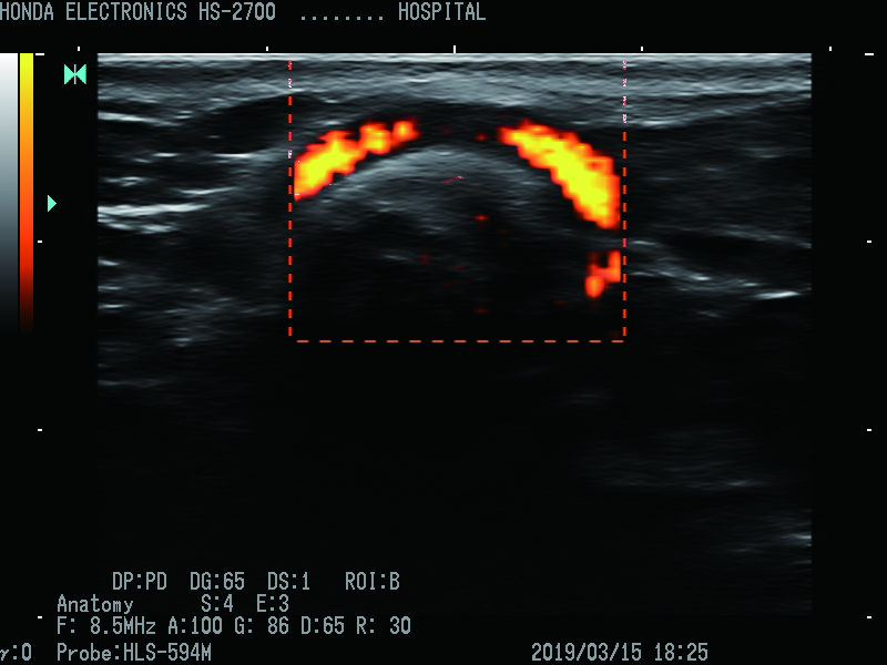
PD(Power Dopller)
Probe:HLS-594M
H-res:Anatomy
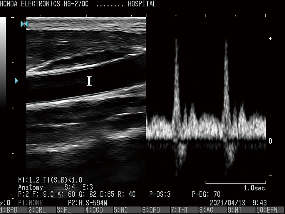
PW(Pulse Doppler)
Probe:HLS-594M
H-res:Anatomy
Box
Probe:HLS-594M
H-res :Anatomy
Vertical
Probe:HLS-594M
H-res :Anatomy

Body mark
A variety of body marks are provided, enabling easy identification and storage of diagnostic area.
*Sample pictures.
Free Download
You can download our product catalog in PDF format from this button.
We hope you will find it useful.
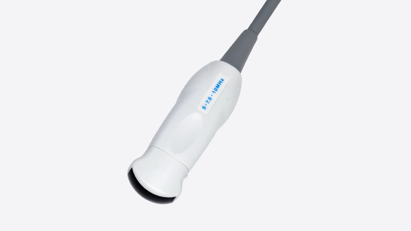
| Type name | HCS-572M |
| Frequency | 9.0/7.5/5.0MHz |
| Radius (sensor part) | 20R |
| Cable length | 2m |
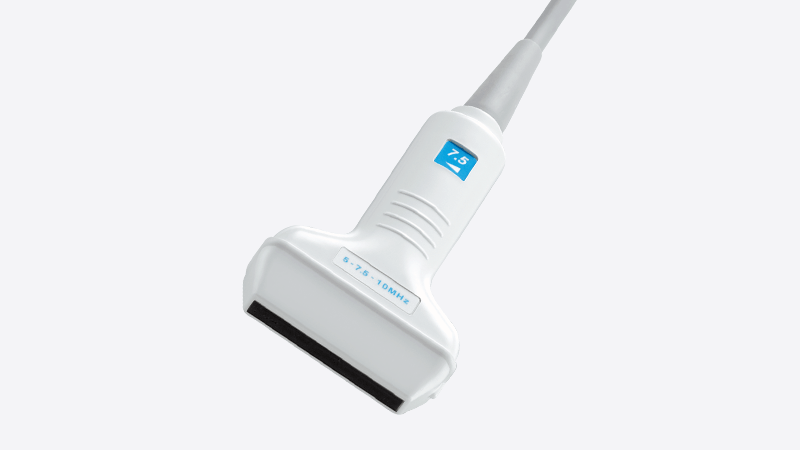
| Type name | HLS-575M |
| Frequency | 10.0/7.5/5.0MHz |
| Width (sensor section) | 50mm |
| Cable length | 2.2m |
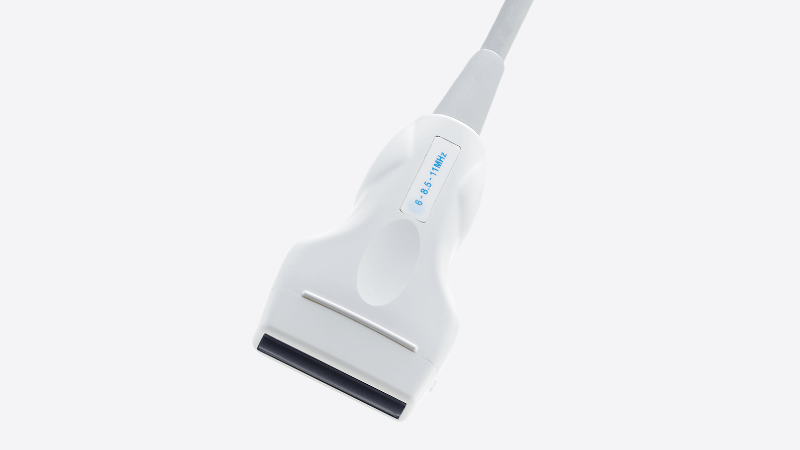
| Type name | HLS-594M |
| Frequency | 11.0/9.0/6.0MHz |
| Width (sensor section) | 40mm |
| Cable length | 2.2m |
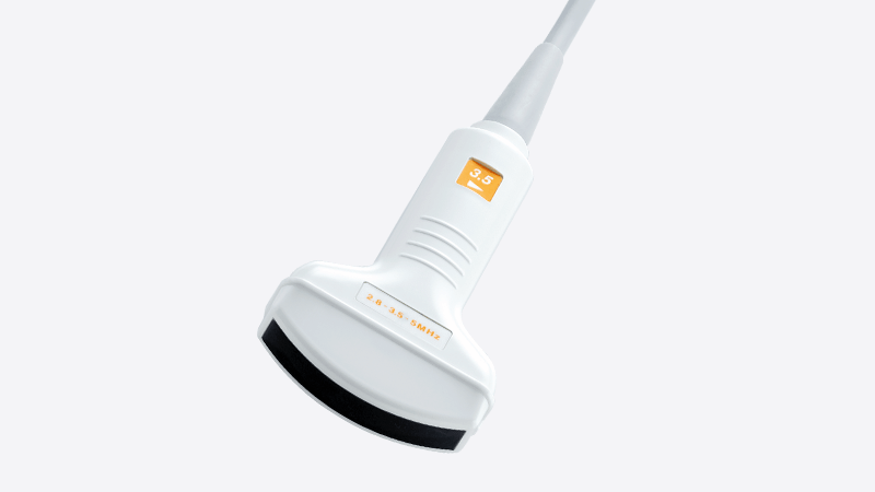
| Type name | HCS-436M |
| Frequency | 5.0/3.5/2.8MHz |
| Radius (sensor part) | 60R |
| Cable length | 2m |
| Type name |
HCS-436MSC *Single crystal probe |
| Frequency | 5.0/3.5/2.8MHz |
| Radius (sensor part) | 60R |
| Cable length | 2m |
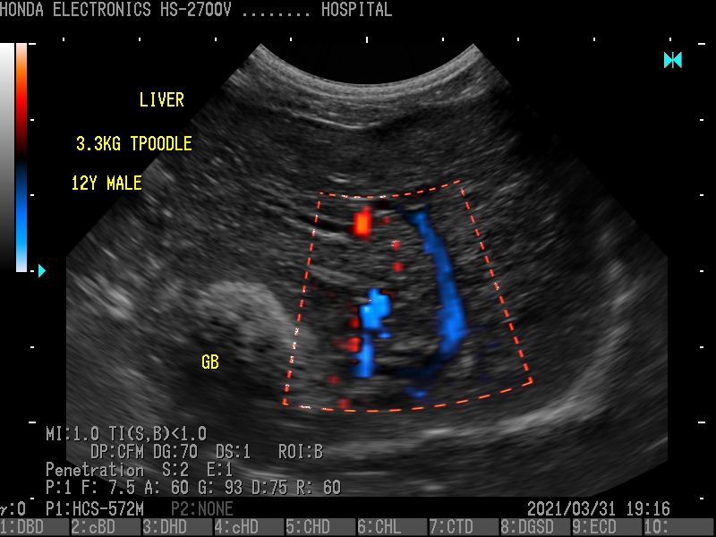
1.Liver (poodle, 12 years old)
Probe:HCS-572M
H-res:Penetration
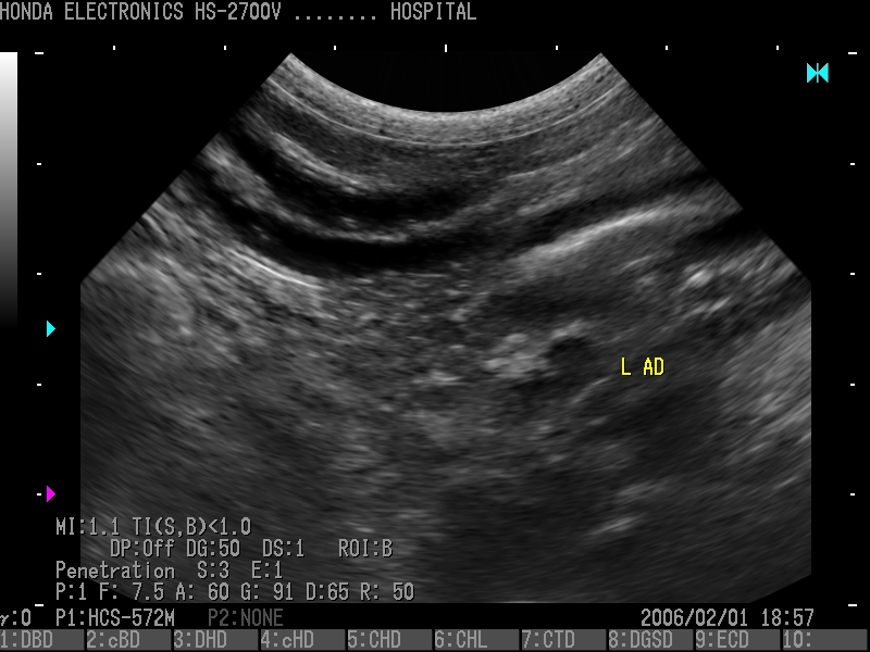
2.Adrenal glands (poodle, 12 years old)
Probe:HCS-572M
H-res : Penetration
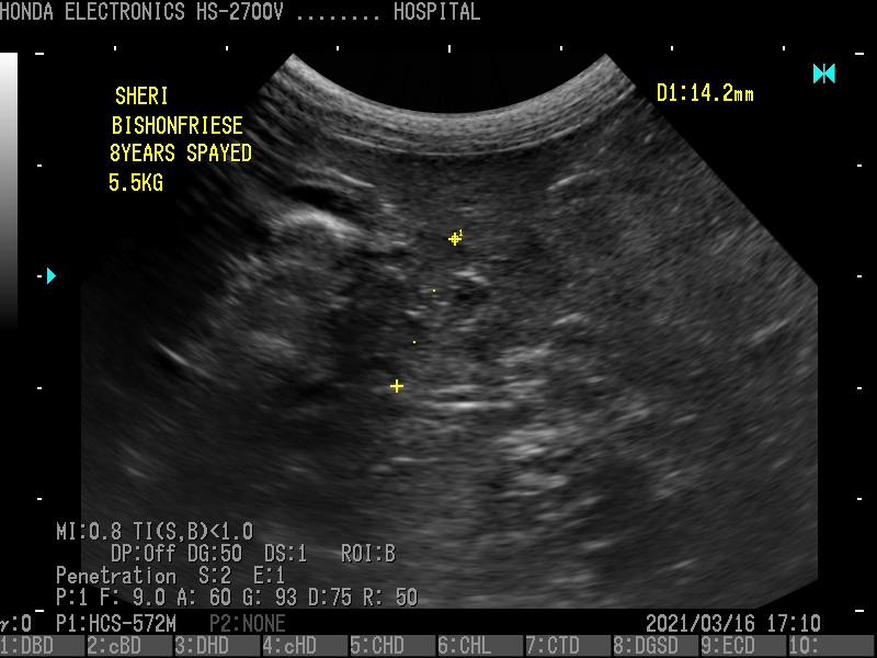
3.Pancreas (Vision Frise, 8 years old)
Probe:HCS-572M
H-res:Penetration
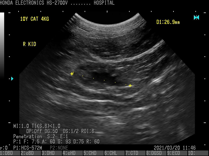
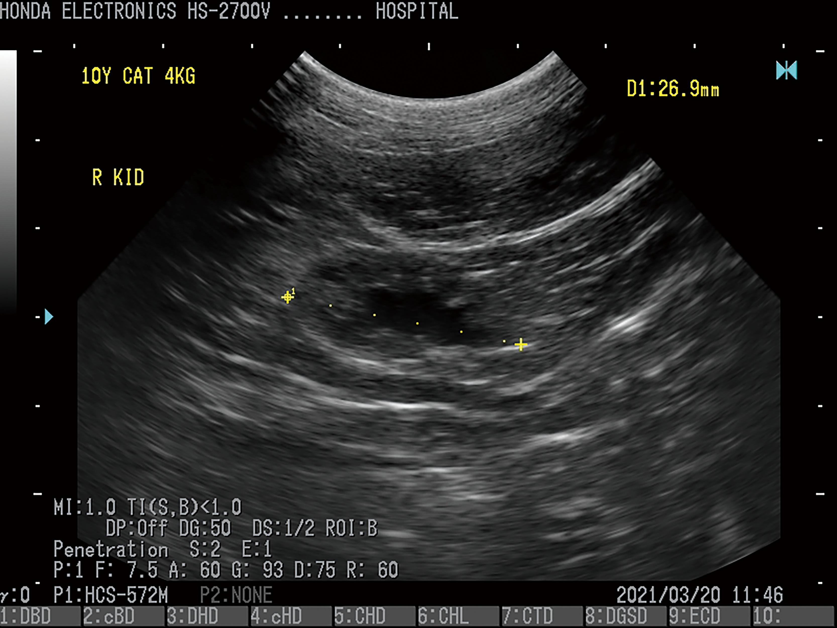
4.Kidney (cat, age 10)
Probe:HCS-572M
H-res : Penetration
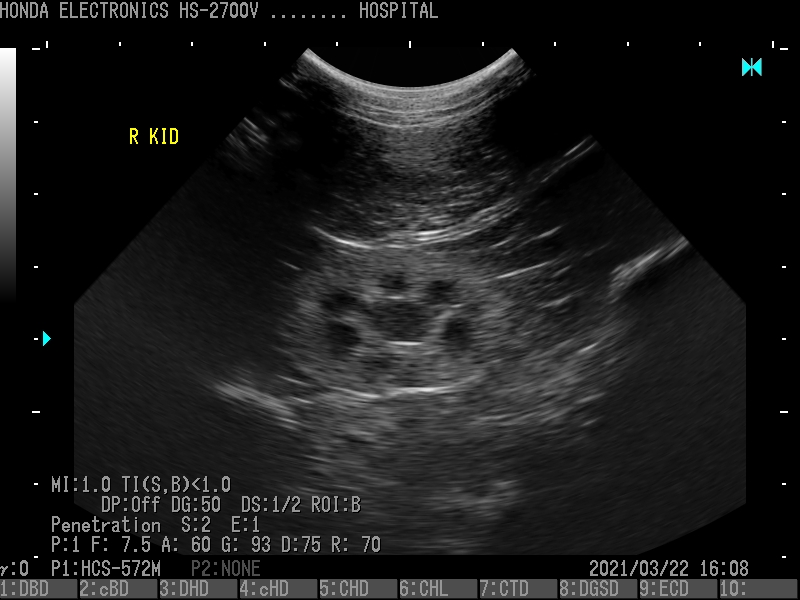
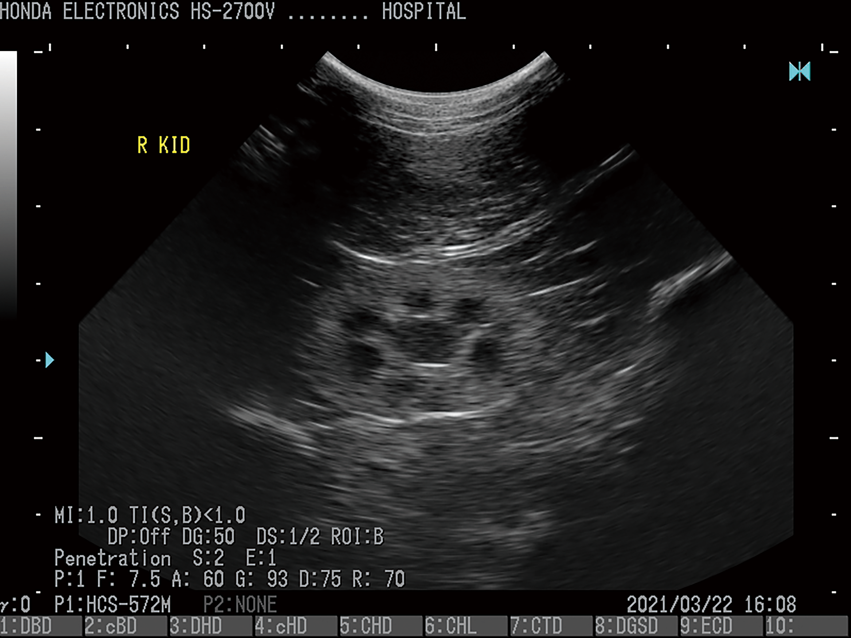
5.Kidney (cat, age unknown)
Probe:HCS-572M
H-res:Penetration
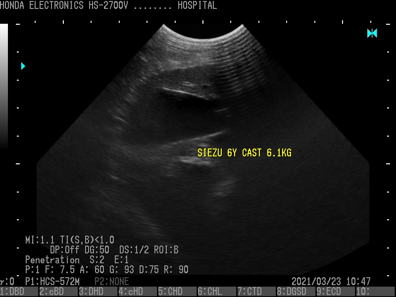
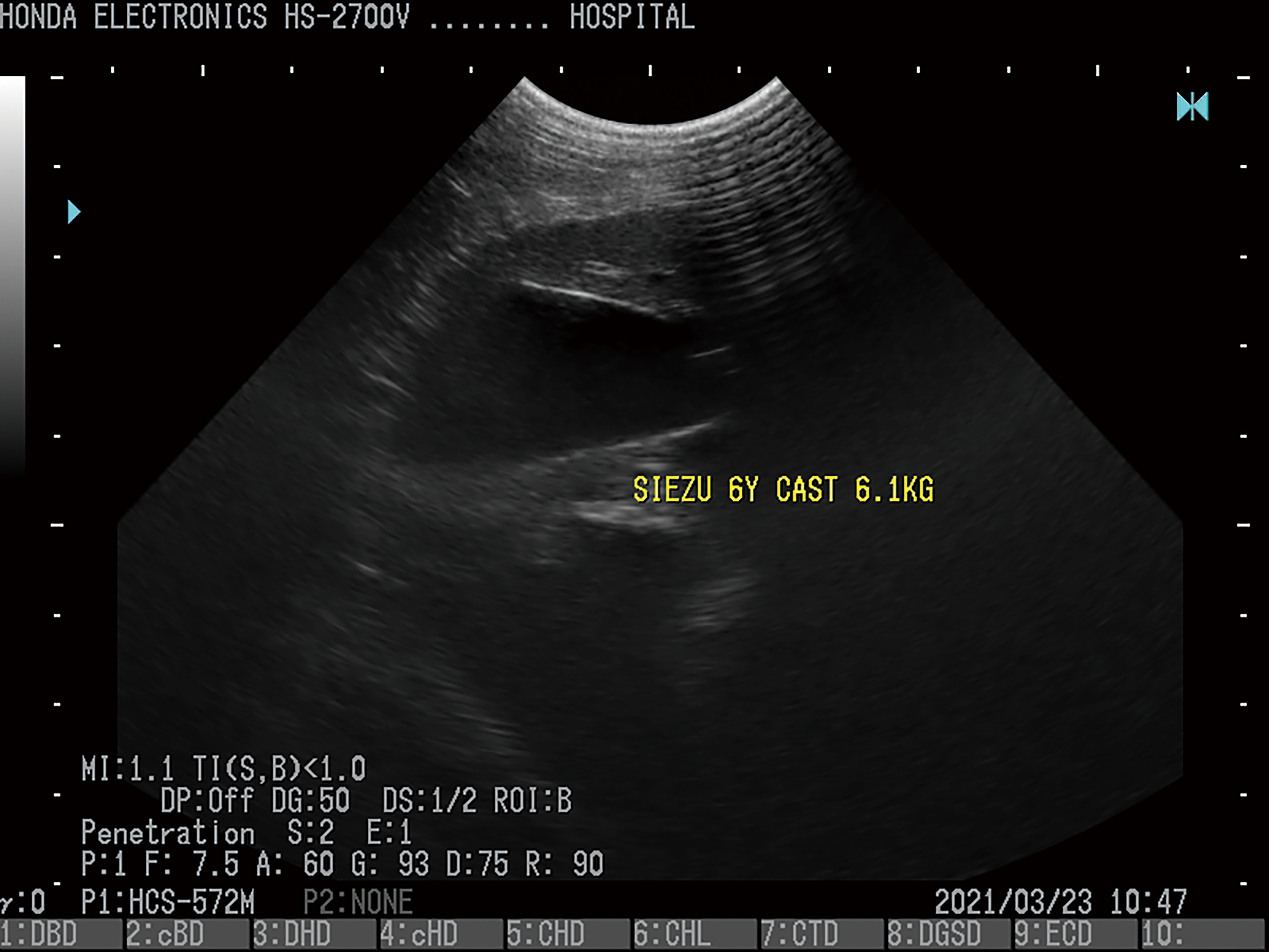
6.Gall bladder (Shih Tzu, 6 years old)
Probe:HCS-572M
H-res : Penetration
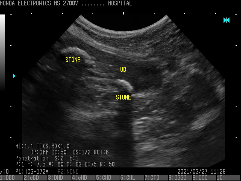
7.Bladder (poodle, 12 years old)
Probe:HCS-572M
H-res:Penetration


